Computational and interferometric imaging
We develop novel forms of quantitative imaging technologies capable of high-throughput, high-resolution and multi-dimensional imaging of various biological and material specimens. To this end, we take multi-modal approaches and combine innovative design of interferometric/non-interferometric optical imaging systems and computational algorithms. Representative technologies are color-coded LED microscopy, smartphone-based multi-contrast microscope via color-multiplexed illumination, and spectral-domain optical coherence phase microscopy.
[1] Lee et al., Color-coded LED microscopy for multi-contrast and quantitative phase-gradient imaging, Biomed. Opt. Express, 6(12), 4912-4922, (2015)
[2] Lee et al., Single-exposure quantitative phase imaging in color-coded LED microscopy, Opt. Express, 25: 8398-8411 (2017)
[3] Jung et al., Smartphone-based multi-contrast microscope using color-multiplexed illumination, 7: 7564, Scientific Reports (2017)
[4] Lee et al., Color-coded LED microscopy for quantitative phase imaging: Implementation and application to sperm motility analysis, Methods, 136: 66-74 (2018)
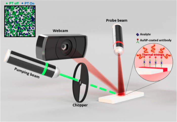
In-vitro diagnostics
A number of molecular diagnostic platforms have been developed to measure abundance of molecules with high-sensitivity, and gain valuable insights into biology at the systems level. Many of these techniques, however, require time-consuming specimen purification and amplification strategies, and may lack the ability for multiplexed measurements desirable in identifying complex diseases, or may not be amenable for easy point-of-care translation. We pursue direct, low-cost, high-throughput molecular assays suitable for point-of-care detection of biomarkers, proteins, and DNA mutation.
[1] Yim et al., Photothermal spectral domain optical coherence reflectometry for direct measurement of hemoglobin concentration of erythrocytes. Biosensors and Bioelectronics. 57, 59-64 (2014).
[2] Kim et al. Capillary-scale direct measurement of hemoglobin concentration of erythrocytes using photothermal angular light scattering, Biosensors and Bioelectronics, 74, 469-475, (2015)
[3] Song et al., Highly sensitive paper-based immunoassay using photothermal laser speckle imaging, Biosensors and Bioelectronics, 117: 385-391 (2018)
In-vivo Microscopy
Optical biopsy refers to methods that utilize the properties of light to obtain a diagnosis of disease during endoscopy and surgery. We explore optical imaging methods and instrumentation that are compatible with endoscopy and surgical procedures. Several technologies have been developed based on our core technologies that include spectral-domain optical coherence tomography (OCT), confocal microscopy, and THz imaging. We also investigate other functional imaging modalities such as fluorescence and nonlinear microscopy to enable comprehensive assessment of biological specimens. Our target clinical applications include oncology, gastroenterology and cardiology.

Tumor discrimination of enhanced green fluorescent protein (eGFP)-transfected human GBM tumorsphere (TS) (eGFP+ GSC-11) tumor-bearing mice (n=4) with TRI and multi-modality imaging. (a) Axial T2-weighted MRI images in living mouse for validation of tumor growth. (b) White light images of the excised brain samples. The tumors were invisible in the white light images as in human malignant gliomas. (c) GFP fluorescence images. (d) Hematoxylin and eosin (H&E) stained image. Both modalities were used for visualization of tumor regions. (e) Optical coherence tomography (OCT) images. These images provide detailed information through the high resolution anatomical structures. Although some regions with reduced scattering may correspond to the tumor region, it is not common feature. (f) TRI images with peak-to-peak amplitude of time-domain signals. Relatively high intensity regions (red) in TRI images are well correlated with real tumor regions that are observed in GFP and H&E stained images. We did not determine the precise threshold value in this preclinical experiment. (g) 5-ALA-induced ppIX fluorescence images. TRI images showed tumor regions more precisely than ppIX fluorescence images. Strong fluorescence in the center of the ppIX images of the mouse brains is emitted not from tumor but the ventricles.
[1] Ji et al., Terahertz reflectometry imaging for low and high grade gliomas, Scientific Reports 6: 36040 (2016)
[2] Triki et al., Intraoperative margin assessment of human breast tissue in optical coherence tomography images using deep neural networks, Computerized Medical Imaging and Graphics, 69: 21-32 (2018)
[3] Kim et al., Spectrally encoded slit confocal microscopy using a wavelength-swept laser, J. Biomed. Opt., 20(3), 036016, (2015).
